Friday, 13 Nov 2026
Speakers
-
 Dr Didier Dietschi, Switzerland
Abstract
This program will focus on modern layering/shading concepts to identify the most efficient approach for single and multiple restorations. Attendees will learn about detailed protocols to achieve high aesthetic results with direct bonding using the” golden standard” 2-layer approach (Natural Layering Concept) with use of effect shades for polychromatic restorations. A special attention will be given to finishing and polishing techniques to achieve optimal primary, secondary and tertiary anatomy with high gloss. Reaching optimal natural & aesthetic integration is no longer an unattainable objective with direct bonding… on the contrary!
Learning objectives
- Identify the indications for direct composite bonding to enhance smile aesthetics in contemporary dental practice
- Determine when full smile enhancement using composite restorations is indicated
- Describe the role of 2D and 3D digital imaging and treatment planning tools.
- Apply fundamental layering and shading concepts to achieve predictable esthetic outcomes with composite materials
Invitation Video
Speakers
-
 Prof Falk Schwendicke, Germany
Abstract
Artificial intelligence (AI) is transforming how clinicians perceive, interpret, and act upon diagnostic information. Yet the step from algorithmic output to informed clinical decision remains complex, requiring not only technical understanding but also critical reflection. This workshop explores how AI can augment human judgment in diagnostic and therapeutic processes, with a particular focus on multimodal data integration—from imaging and sensor signals to clinical and behavioral information.
Participants will gain hands-on experience with an AI-supported diagnostic tool, experimenting with real-world datasets and multimodal sensor inputs. Through guided exercises and group discussions, we will examine how AI-driven insights can inform, challenge, or refine clinical decisions. Beyond demonstrating current capabilities, the workshop will address limitations, transparency, and ethical dimensions of AI-assisted decision-making, as well as the need for what is now called „data literacy“.
By the end of the session, attendees will have developed a practical and conceptual understanding of how AI systems process complex diagnostic information and how clinicians can critically engage with these outputs to improve care quality and patient outcomes.
Speakers
-
 Dr Rupert Austin, UK
Abstract
Erosive tooth wear and dentine hypersensitivity are increasingly common clinical challenges, often presenting together and frequently driven by overlapping dietary, behavioural, medical, and restorative risk factors. This lecture will explore a practical, prevention-led approach to identifying, diagnosing, and managing erosive tooth wear and associated sensitivity in contemporary dental practice.
The session will link clinical risk assessment with patient-centred communication, early intervention, and long-term maintenance. It will review how to distinguish erosive tooth wear from other patterns of tooth surface loss, how to identify modifiable aetiological factors, and how to integrate behavioural advice, biofilm control, fluoride-based strategies, desensitising technologies, and restorative decision-making. Particular emphasis will be placed on minimally invasive management, protection of dentine and pulpal health, and maintaining oral health-related quality of life.
By the end of the lecture, delegates should be able to move beyond symptomatic relief alone and adopt a structured approach that combines prevention, sensitivity control, restorative planning, and maintenance for patients with erosive tooth wear. This aligns with the prosthodontic emphasis on risk assessment, prevention, behavioural advice, treatment planning, and maintenance in erosive tooth wear.
Learning Objectives
- Recognise the clinical presentation and progression of erosive tooth wear and its relationship with dentine hypersensitivity.
- Differentiate erosive tooth wear from attrition, abrasion, abfraction, caries-related tissue loss, and post-restorative sensitivity.
- Identify dietary, behavioural, medical, salivary, occupational, and restorative risk factors that contribute to erosive tooth wear.
- Apply a structured chairside risk assessment to guide prevention, monitoring, sensitivity control, and restorative decision-making.
- Select appropriate preventive and non-invasive strategies, including behavioural modification, fluoride-based protection, and desensitising technologies.
- Integrate minimally invasive restorative principles when tooth wear affects function, aesthetics, pulpal health, or quality of life.
- Develop maintenance and recall strategies for patients at ongoing risk of erosive tooth wear and recurrent dentine hypersensitivity.
Speakers
-
 Dr Pokpong Amornvit, Thailand
Abstract
Contemporary dental treatment planning increasingly emphasizes a “begin with face” approach, recognizing the face as the primary reference for achieving optimal esthetic and functional outcomes. Traditional workflows often rely solely on intraoral landmarks, which may limit the predictability and harmony of the final result. In contrast, integrating facial analysis into treatment planning enables clinicians to align dental structures with overall facial proportions, enhancing both precision and patient-specific customization.
With the rapid advancement of digital dentistry, a wide range of technologies now support this face-driven workflow. Tools such as facial scanners, intraoral scanners, and cone beam computed tomography (CBCT) facilitate comprehensive data acquisition, while advanced software platforms enable integration, simulation, and design across multiple domains. These technologies improve diagnostic accuracy, streamline communication, and enhance treatment predictability.
This lecture provides a systematic overview of various digital dental tools and compares their capabilities in capturing measurements, analyzing anatomical relationships, and supporting treatment design. Emphasis is placed on how each modality contributes uniquely to the workflow, from facial reference acquisition to prosthetic planning, implant positioning, and orthodontic simulation.
By demonstrating practical applications across restorative dentistry, implantology, and clear aligner therapy, this session highlights how a face-driven digital workflow can be effectively implemented in daily clinical practice. The objective is to provide clinicians with a clear understanding of which tools to use, what parameters they can measure, and how they can be integrated to achieve precise, predictable, and esthetically driven outcomes.
Learning Objectives
- Discuss the concept of “begin with face” and its importance in modern dental treatment planning.
- Explain how facial reference integration improves predictability and precision in dental outcomes.
- Describe the roles of key digital dental technologies, including facial scanners, intraoral scanners, and CBCT.
- Compare the capabilities of different digital tools in measurement, analysis, and treatment design.
- Demonstrate how to integrate multiple digital datasets into a cohesive, face-driven workflow.
- Apply face-driven digital planning principles to restorative, implant, and orthodontic cases.
Speakers
-
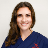 Dr Larissa Steigmann, Germany
-
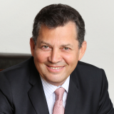 Dr Marius Steigmann, Germany
Abstract
Achieving optimal aesthetic outcomes in implant dentistry involves not only the successful integration of the implant with the surrounding bone but also the harmonious integration of the peri-implant soft tissues. Soft tissue management in the aesthetic zone plays a crucial role in creating natural-looking restored implants and satisfying patient expectations. Successful soft tissue management requires meticulous planning, precise surgical techniques, and attention to detail. It also necessitates an understanding of the biological principles governing soft tissue healing and the interplay between the soft tissue and the underlying bone, creating an aesthetically pleasing emergence profile that mimics natural teeth.
Incision, flap design and suturing techniques are indication specific. E.g., oral surgery, periodontal surgery and endodontic surgery require different soft tissue manipulation dependent on the goal of the surgery. Hence, clear rules are to be respected, according to the objective of the surgery, phenotype of the patient, location in the oral cavity, size of the surgical site – hence, different approaches are to be chosen. Every surgery when performed can have several complications including flap opening, recession, papilla loss, tissue necrosis (etc.). These are the consequences if the focus is single sided with the only goal to reach primary closure without paying attention to the collateral damage of the surgical intervention. The lecture will focus on decision factors prior to the surgery to avoid aesthetic complications.
Learning Objectives
- Understand the biological principles governing peri-implant soft tissue healing and their impact on aesthetic outcomes.
- Identify indication-specific incision, flap design, and suturing techniques based on surgical objectives and clinical scenarios.
- Evaluate patient- and site-related factors (soft tissue phenotype, location, and defect size) influencing soft tissue management decisions.
- Recognize common aesthetic complications associated with improper soft tissue handling and their underlying causes.
- Apply pre-surgical decision-making strategies to minimize soft tissue trauma and prevent aesthetic failures in the implant aesthetic zone.
Speakers
-
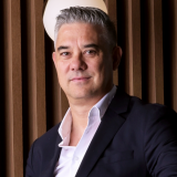 Dr Daniel Baketić, Croatia
Abstract
3D printing is rapidly expanding beyond models and provisional restorations, offering new possibilities for minimally invasive restorative dentistry. This lecture presents a practical digital workflow combining biomimetic principles, adhesive protocols, intraoral scanning, CAD design, and chairside 3D printing.
Through clinical cases, the presentation will demonstrate how 3D printing can be integrated into indirect restorative procedures, with a focus on definitive restorations, workflow efficiency, material selection, post-processing, and clinical application.
The aim is to demonstrate how additive manufacturing can be incorporated into biologically driven, minimally invasive protocols while improving predictability, efficiency, and control over the restorative workflow.
Learning Objectives
- Explain the biomechanical principles that guide the decision between direct and indirect restorative approaches while preserving healthy tooth structure.
- Discuss how minimally invasive concepts such as cavity design optimization, immediate dentin sealing, and posterior indirect adhesive restorations can improve the predictability of restorative treatment.
- Describe a complete digital restorative workflow, from tooth preparation and intraoral scanning through digital design, 3D printing, post-processing, and adhesive cementation.
- Evaluate the clinical factors that influence intraoral scanning accuracy, material selection, and the performance of 3D-printed definitive restorations.
- Demonstrate how digital technologies can support more conservative clinical decision-making, enabling efficient same-day treatment while reducing unnecessary removal of healthy tooth structure.
Speakers
-
 Dr Thamer Theeb, Jordan
Abstract
Adhesive dentistry has transformed restorative treatment by allowing clinicians to preserve tooth structure while achieving predictable functional and aesthetic outcomes. This lecture presents a stepwise approach to adhesive rehabilitation, beginning with simple direct restorations and progressing to complex full-mouth cases. The presentation will explore case selection, diagnosis, treatment planning, occlusal considerations, material selection, bonding protocols, and the integration of direct and indirect restorations. Particular emphasis will be placed on minimally invasive strategies, including additive wax-ups, mock-ups, vertical dimension modification, partial-coverage restorations, and bonded ceramic rehabilitation. Through clinical cases of increasing complexity, the lecture will demonstrate how the same fundamental adhesive principles can be applied from a single tooth to a complete functional and aesthetic reconstruction. Common complications, limitations, and decision-making challenges will also be discussed. The objective is to provide clinicians with a practical framework for planning and executing adhesive rehabilitations predictably, conservatively, and with confidence.
Learning Objectives
- Discuss the biological, functional, and aesthetic principles of adhesive rehabilitation.
- Explain the diagnostic and treatment-planning process for cases ranging from single-tooth restorations to full-mouth rehabilitation.
- Demonstrate appropriate case selection and clinical decision-making for direct and indirect adhesive restorations.
- Describe minimally invasive strategies for preserving tooth structure, including additive wax-ups, mock-ups, and partial-coverage restorations.
- Explain the principles of vertical dimension modification and occlusal management in complex rehabilitations.
- Compare restorative materials and bonding protocols according to clinical indications and substrate conditions.
- Discuss common complications, limitations, and maintenance considerations associated with adhesive rehabilitation.
- Apply a predictable stepwise workflow for integrating direct and indirect restorations in full-mouth cases.
Speakers
-
 Dr Roberto Turrini, Italy
Abstract
Tooth discoloration represents a complex challenge in aesthetic dentistry, resulting from a wide spectrum of intrinsic and extrinsic factors, including enamel defects and alterations related to vital and non-vital teeth. A thorough understanding of the biological mechanisms governing color expression in enamel and dentin is essential for achieving predictable and durable aesthetic outcomes.This lecture analyzes the etiology of tooth discoloration, with particular emphasis on enamel defects, chromophore dynamics, and the structural differences between vital and non-vital teeth. A structured diagnostic workflow is presented to support accurate classification and appropriate treatment planning.Contemporary aesthetic strategies are discussed, ranging from minimally invasive whitening protocols to restorative and prosthetic solutions, guided by optical principles, material science, and tissue preservation. Clinical cases demonstrate how biological insight enables effective aesthetic control, even in complex discoloration scenarios.By integrating biology, diagnosis, and modern aesthetic techniques, this presentation provides clinicians with a scientifically sound framework for managing tooth discoloration with precision, consistency, and respect for dental tissues.
Learning Objectives
- Discuss the biological mechanisms of tooth discoloration, including enamel defects in vital and non-vital teeth.
- Explain the diagnostic criteria for differentiating intrinsic and extrinsic discolorations.
- Discuss contemporary aesthetic strategies for the management of tooth discoloration.
- Demonstrate material selection based on optical properties and biological principles.
- Demonstrate a structured, biologically driven approach to aesthetic control of complex discoloration cases.
Speakers
-
 Prof Dr Esra Uzer Celik, Türkiye
Abstract
Achieving natural-looking smiles is one of the most challenging goals in contemporary aesthetic dentistry. Patients increasingly demand restorations that harmonize with natural dentition in terms of color, translucency, surface texture, and anatomical form. To meet these expectations, clinicians must combine scientific knowledge of dental materials with a strong understanding of dental anatomy and artistic skills.
This lecture focuses on the key principles required to create highly aesthetic and natural-appearing restorations in daily clinical practice. Special emphasis will be placed on the importance of proper diagnosis, shade selection, layering strategies, anatomical reconstruction, and surface texture reproduction. Participants will learn how small details such as line angles, macro- and micro-texture, and polishing protocols significantly influence the final aesthetic outcome.
Through clinical cases and practical tips, the lecture will demonstrate predictable approaches that simplify complex aesthetic procedures while maintaining high clinical standards. The goal is to help clinicians develop a systematic workflow for achieving natural results efficiently and consistently.
By understanding the artistic and scientific aspects of smile creation, clinicians can improve both the aesthetic quality and long-term success of their restorative treatments.
Learning Objectives
After attending this lecture, participants will be able to:
- Discuss the key principles involved in creating natural-looking smiles in aesthetic dentistry.
- Explain the role of dental anatomy, surface texture, and morphology in achieving natural aesthetics.
- Demonstrate how layering strategies influence color depth, translucency, and overall smile appearance.
- Explain how line angles, macro-texture, and micro-texture contribute to the perception of natural teeth.
- Discuss simplified clinical strategies for predictable aesthetic outcomes in daily practice.
- Demonstrate practical techniques for improving finishing and polishing to enhance surface texture and optical properties.
No programs of this type on this day
Saturday, 14 Nov 2026
Speakers
-
Dr Roberto Turrini, Italy
Abstract
Nowadays, minimally invasive prosthetic procedures transcend traditional full-crown dentistry. By reducing tooth reduction and using adhesive cementation, these techniques maintain pulp vitality, maximise fracture resistance and achieve long-lasting, high-definition aesthetics and function. This presentation is distilled into a complete, step-by-step workflow: proper diagnosis, guided preparation, ultra-precise impressions or digital scans, and safe adhesive cementation. Case reports — from single veneers to full-mouth adhesive restorations — demonstrate how a broad palette of contemporary materials and techniques meet biological, functional, and cosmetic demands. Participants will gain ready-to-apply clinical tips and discover how emerging digital trends integrate with classical protocols to streamline chair time, refine outcomes, and simplify maintenance, focusing on all modern restorative alternatives, including difficult cases such as discolorations and worn dentition.
Learning objectives
- Discuss the biological, functional, and aesthetic principles of modern adhesive prosthetic dentistry.
- Explain diagnostic criteria and case selection for minimally invasive adhesive restorations.
- Demonstrate step-by-step workflows for preparation, impressions or digital scans, and adhesive cementation.
- Explain the clinical use of contemporary adhesive materials to enhance longevity and aesthetics.
- Discuss the integration of digital technologies into adhesive prosthetic protocols.
Speakers
-
Dr Pokpong Amornvit, Thailand
Abstract
Conventional implant surgery typically involves flap elevation to allow direct visualization of the underlying bone. Although effective, this approach may lead to increased postoperative pain, swelling, and delayed healing. With the advancement of digital dentistry, minimally invasive techniques—particularly flapless implant surgery—have become increasingly achievable through the use of digital surgical guides.
Modern workflows integrate intraoral scanning and cone beam computed tomography (CBCT) to capture both soft tissue and bone anatomy. These datasets are merged within planning software to create patient-specific surgical guides, enabling precise three-dimensional implant placement while minimizing surgical trauma. As a result, patients benefit from reduced discomfort and accelerated recovery.
In clinical practice, implant placement has traditionally relied on the experience and judgment of highly skilled clinicians—referred to here as “GuruGuide.” While clinical expertise remains invaluable, it does not always ensure consistency or precision, particularly in complex or high-volume cases where variability and human error may occur. In contrast, digital surgical guides provide a standardized and reproducible approach that enhances accuracy and reduces operator-dependent variability.
This lecture will compare the concepts of “GuruGuide” and Digital Surgical Guide, highlighting differences in predictability, workflow, and clinical outcomes. Various clinical scenarios, ranging from single-tooth implants to full-arch rehabilitation, will be presented to illustrate appropriate applications and limitations. The session aims to provide clinicians with a clear understanding of how digital guidance can complement clinical expertise to achieve more precise, minimally invasive, and predictable implant treatment outcomes.
Learning Objectives
- Discuss the principles of minimally invasive implant surgery and the role of flapless techniques.
- Explain the integration of intraoral scanning and CBCT in digital implant planning.
- Compare Digital Guide and GuruGuide systems in terms of design, workflow, and clinical application.
- Demonstrate the step-by-step workflow for creating and utilizing surgical guides in implant surgery.
- Describe the indications and limitations of guided implant surgery in single-tooth and full-arch cases.
- Apply digital guided surgery concepts to improve precision, reduce patient morbidity, and enhance clinical outcomes.
Speakers
-
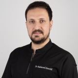 Dr Mahmood Fawzi Chawshli, Iraq
Abstract
Full-mouth rehabilitation is a complex clinical procedure that requires careful integration of aesthetics, function, occlusion, and biological considerations. The development of digital dentistry has significantly improved the predictability, efficiency, and communication involved in comprehensive restorative treatment. Zirconia, with its high strength, favourable aesthetic properties, and biocompatibility, has become an important material for extensive fixed prosthetic rehabilitation.
This case describes the full-mouth rehabilitation of a patient with multiple compromised teeth, altered occlusal relationships, and aesthetic concerns using [monolithic / multilayered] zirconia restorations. A complete digital workflow was implemented from diagnosis to definitive prosthetic delivery. Initial records included intraoral digital scanning, standardised clinical photography, and digital radiographic assessment. The occlusal relationship, vertical dimension, tooth proportions, and smile characteristics were analysed digitally. A virtual diagnostic wax-up was then designed to establish the proposed tooth morphology, occlusal scheme, and aesthetic parameters.
The digital design was transferred to the clinical setting through digitally fabricated provisional restorations, allowing evaluation of phonetics, function, occlusion, facial support, and patient acceptance. Following clinical verification and the necessary modifications, the definitive zirconia restorations were designed and manufactured using computer-aided design and computer-aided manufacturing (CAD/CAM) technology. The restorations were assessed clinically for marginal adaptation, proximal contacts, occlusion, and aesthetics before final cementation.
The integration of zirconia restorations with a complete digital workflow provides a predictable and efficient approach to full-mouth rehabilitation. Digital scanning, virtual treatment planning, CAD/CAM fabrication, and digitally guided provisionalisation support improved communication, precise restorative design, and effective control of aesthetic and functional parameters. This approach may reduce clinical adjustments while enhancing patient comfort and treatment predictability. Successful outcomes nevertheless remain dependent on accurate diagnosis, appropriate occlusal planning, meticulous clinical execution, and careful selection of the zirconia type and the bonding or cementation protocol.
Learning Objectives
- Describe the principles of full-mouth rehabilitation.
- Explain digital workflows in restorative treatment.
- Evaluate esthetic and occlusal parameters digitally.
- Discuss zirconia selection for full-mouth restorations.
- Demonstrate CAD/CAM planning and restorative delivery.
Speakers
-
 Dr Marcus Dagnelid, Sweden
Abstract
This lecture will explore modern digital implant workflows, focusing on techniques and protocols that enable predictable outcomes through the use of intraoral scanning. Emphasis will be placed on digital impression from single to fully edentulous patients, supported by current research and clinical applications. Participants will gain insights into how digital technologies can enhance precision, improve clinical efficiency, and increase treatment predictability in everyday practice. The lecture presents elements such as healing abutment concepts, definitive abutment solutions and state-of-the-art bridge concepts.
Speakers
-
 Dr Maurizio Martini, Italy
Abstract
Coronectomy was first described by Ecuyer and Debien in 1984 as a procedure for partial extraction of third molars. Most recently, it has been investigated as an alternative to traditional surgical extraction of third molars, particularly for those with an increased risk of damage to the inferior alveolar nerve (IAN). Several studies have demonstrated that coronectomy does significantly decrease the risk of iatrogenic injury to the IAN, with some studies also suggesting a lower complication rate. Knowing how to perform this kind of procedure puts the dentist in the position to offer the patient a safer and predictable procedure when it comes to the extraction of risky wisdom teeth.
Learning objective
- Radiological signs of risk of damaging the IAN during wisdom teeth evaluation
- Evaluation of coronectomy as alternative to the surgical extraction of wisdom teeth
- Surgical technique of coronectomy: what to do
- Importance of the instrments for the right and safer surgical procedure.
Speakers
-
Dr Maurizio Martini, Italy
Abstract
Prevention of peri-implantitis in the implant therapy is a key for the long term success of the entire restoration. Choose the right features of your implants together with a proper management of the surgery is pivotal to guarantee a perfect start for your implant rehabilitations, while professional maintenance and patient collaboration will do the rest in other to keep your implants healthy and efficient for many years.
Learning Objectives
By the end of this lecture, participants will be able to:
- Identify key risk factors for peri-implant disease in patients with a history of periodontitis.
- Explain the importance of appropriate implant selection and surgical management for long-term success.
- Discuss strategies for preventing peri-implantitis before and after implant placement.
- Develop effective professional maintenance and follow-up protocols for implant patients.
- Educate patients on oral hygiene and long-term compliance to maintain peri-implant health.
Speakers
-
 Dr Jan Paulics, Sweden
Abstract
Artificial intelligence is expanding the clinical value of intraoral scanning beyond documentation and treatment planning. By analyzing scan data and highlighting potential oral health findings, AI-assisted tools can support clinicians in identifying surface and proximal caries, plaque, gingival recession, and tooth wear.
This lecture explores how visual overlays, structured assessments, and longitudinal scan comparisons can contribute to more consistent clinical evaluations and data-informed treatment decisions. Sequential scans may help clinicians monitor changes over time, assess disease progression, evaluate treatment outcomes, and communicate the importance of preventive care.
The presentation will also demonstrate how complex clinical information can be transformed into clear and accessible visual explanations for patients. Patient-facing digital tools can reinforce conversations in the dental practice, improve understanding, and encourage greater participation in treatment and prevention.
Through clinical examples and practical workflows, participants will gain insight into how AI-assisted intraoral analysis can enhance diagnostic confidence, strengthen patient communication, and support a more preventive, personalized, and patient-centered approach to oral healthcare. The lecture will also address the importance of responsible implementation, data quality, and continued clinical oversight when incorporating AI into everyday practice.
Learning Objectives
- Understand the foundations of clinical AI: Explain how artificial intelligence can be trained on large datasets of intraoral scans to support consistent identification of oral health findings.
- Recognize AI-assisted clinical findings: Explore the identification and visualization of surface caries, proximal caries, plaque, gingival recession, and tooth wear.
- Apply longitudinal monitoring: Understand how sequential intraoral scans can be compared to monitor changes, disease progression, and treatment outcomes over time.
- Strengthen patient communication: Use intuitive visual overlays and patient-facing digital tools to explain clinical findings and encourage greater patient understanding, engagement, and participation.
- Integrate AI responsibly into clinical practice: Recognize AI as a decision-support tool that complements, rather than replaces, professional clinical judgment.
Supported by

Speakers
-
 Dr Shaista Siddiqui, UK
-
 Dr Gilberte Rechdan, Lebanon
Abstract
Discover how clear aligner therapy can enhance patient care and support practice growth. Through clinical case examples and digital workflows, learn how to improve diagnosis, treatment planning, patient communication, and case acceptance while incorporating aligner therapy seamlessly into everyday practice.
Learning outcomes
- Identify opportunities to incorporate clear aligner therapy into daily practice.
- Understand how digital workflows support diagnosis and treatment planning.
- Explore clinical cases demonstrating predictable outcomes.
- Improve patient communication and case acceptance using digital tools.
- Gain practical strategies to introduce and grow aligner therapy in your practice.
Speakers
-
Prof Falk Schwendicke, Germany
Abstract
White enamel lesions are common in clinical practice but may have different etiologies, characteristics, and treatment requirements. Distinguishing early carious white spot lesions from developmental enamel defects such as Molar Incisor Hypomineralization (MIH) is therefore essential for appropriate treatment planning.
Designed for a general dental audience, this lecture provides a practical overview of white spot lesions and MIH, moving from diagnosis and differential diagnosis to treatment selection and long-term management. Participants will be introduced to a structured clinical pathway that integrates prevention, remineralization, resin infiltration, aesthetic management, and restorative treatment according to lesion characteristics and patient needs.
Particular emphasis will be placed on combining treatment modalities and how to come to reliable aesthetic results with the least invasive approaches.
Learning objectives
By the end of this lecture, participants will be able to:
- Explain the key clinical features and differential diagnosis of white spot lesions and MIH.
- Discuss how lesion characteristics, severity, and patient factors influence treatment selection.
- Apply a structured clinical pathway for the management of MIH and other white spot lesions.
- Demonstrate appropriate case selection for preventive, remineralizing, infiltrative, and restorative approaches.
- Explain how different treatment modalities can be combined to achieve functional and aesthetic outcomes.
- Discuss strategies for reducing hypersensitivity and improving patient comfort during treatment.
- Evaluate clinical cases and formulate individualized treatment plans that support predictable, long-term outcomes.
Speakers
-
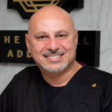 Prof Tarek Salah Morsi, Egypt
Abstract
Current intraoral scanning technologies primarily capture static anatomical data, while functional occlusion remains largely inferred. Emerging innovations aim to bridge this gap through real-time functional scanning, combining motion tracking, AI, and advanced sensors. This lecture explores the evolution of scanning technologies toward dynamic occlusion capture, discussing their potential to redefine diagnosis, prosthetic design, and implant success prediction.
Learning Objectives
- Explain the Evolution of intraoral scanning
- Understand the limitations of static intraoral scanning
- Explore emerging technologies in real-time functional scanning
- Evaluate clinical applications of dynamic occlusion capture
- Anticipate future developments in digital dentistry
Speakers
-
 Dr Fadi Khuffash, UAE
Abstract
Light curing remains essential for the success of resin-based restorations. Advances from halogen to LED technologies have improved efficiency, with dual-wavelength (polywave) LEDs providing broader spectral coverage. Unlike single-peak LEDs targeting camphorquinone, polywave units also activate alternative photoinitiators such as PPD, TPO, and Ivocerin, ensuring reliable polymerization in esthetic and high-opacity composites. Dual-cure systems further enhance outcomes in areas with limited light transmission, such as deep cavities and indirect restorations. This lecture reviews these technologies and their clinical implications for predictable, durable restorations.
Learning Objectives
- Trace the evolution of dental light curing technologies through the different generations, recognizing their clinical implications.
- Differentiate between halogen and LED curing lights in terms of energy output, wavelength spectrum, efficiency, and clinical effectiveness.
- Evaluate the benefits and limitations of dual-cure systems in situations such as deep cavities, fiber posts, and indirect restorations.
- Identify factors affecting depth of cure and polymerization quality to reduce restoration failures and post-operative sensitivity.
- Apply evidence-based guidelines for selecting and using curing lights to achieve predictable and durable restorative outcomes.
Speakers
-
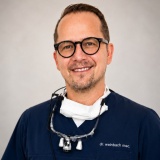 Dr Christoph Weinbach, Germany
Abstract
As patient expectations for highly esthetic, biocompatible, and metal-free treatment solutions continue to grow, zirconia implants are gaining increasing attention in modern implant dentistry. This lecture is designed specifically for dental professionals seeking an evidence-based understanding of zirconia implant therapy and its integration into contemporary digital workflows.
The lecture will review the scientific foundations of zirconia implants, focusing on material properties, osseointegration, peri-implant soft tissue behavior, and long-term clinical performance.
Special attention will be given to the simplicity and efficiency of the SDS Swiss Dental Solutions implant system, highlighting both the surgical and prosthodontic aspects of the workflow. Participants will gain practical insight into streamlined treatment protocols, minimally invasive surgical approaches, and simplified restorative procedures that support predictable esthetic and functional outcomes in daily clinical practice.
A significant part of the lecture will focus on extensive clinical case presentations from the author’s daily practice, with particular emphasis on treatments in the esthetic zone. Immediate implantation protocols and their clinical advantages will also be emphasized, particularly in relation to preserving hard and soft tissue architecture, reducing treatment time, and optimizing esthetic outcomes. Through real-world cases involving single-tooth, partially edentulous, and full-arch rehabilitations, participants will gain practical insight into treatment planning, immediate implant placement, soft tissue management, prosthetic integration, and long-term esthetic outcomes when using zirconia implants in highly demanding clinical situations.
In addition to scientific evidence, the lecture will provide practical clinical perspectives relevant to everyday dentistry, including patient selection, surgical and prosthetic protocols, restorative workflows, maintenance strategies, and interdisciplinary treatment planning.
By combining science, digital innovation, and real-world clinical experience, this session aims to provide dentists, implantologists, prosthodontists, and restorative clinicians with practical knowledge and confidence in incorporating zirconia implants into modern esthetic and biologically driven dental practice."
Learning objectives
- Participants will gain the ability to discuss the scientific foundations of zirconia implants, including material properties, osseointegration, peri-implant soft tissue behavior, and long-term clinical performance.
- Participants will be able to explain the indications, advantages, and limitations of zirconia implants compared with conventional titanium implant systems in modern dental practice.
- Through clinical demonstrations and workflows, participants will better understand and demonstrate the simplicity and efficiency of implant system in both surgical and prosthodontic applications.
- Participants will learn to evaluate immediate implantation protocols in the esthetic zone, including techniques for preserving hard and soft tissue architecture and optimizing esthetic outcomes.
- By reviewing extensive clinical cases, participants will develop the ability to analyze treatment planning, prosthetic integration, and interdisciplinary decision-making in single-tooth, partially edentulous, and full-arch zirconia implant rehabilitations.
No programs of this type on this day
Chairperson
Thank you to our chairperson for their support during the event.